It Travels Inert. Then It Meets a Cell.
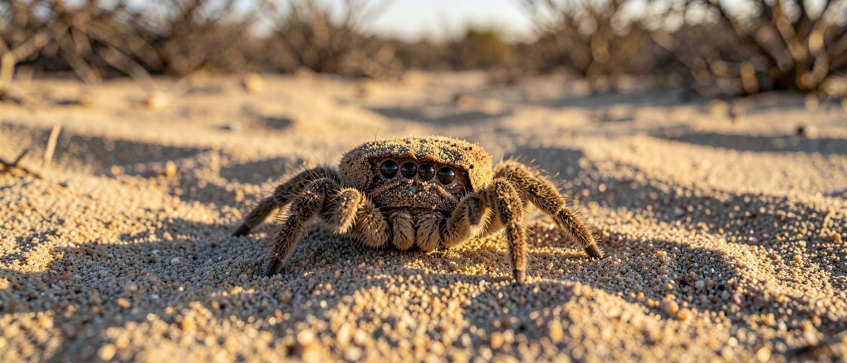
The venom enzyme that makes a Chilean six-eyed sand spider (Sicarius levii) dangerous does essentially nothing while it moves through a body, no membrane disruption, no tissue attack. It circulates inert until it physically contacts a cell surface, and then it switches on.
That activation-on-contact finding is the center of a paper Alexandra Sundman, Greta Binford, William Montfort, and Matthew Cordes published in the Proceedings of the National Academy of Sciences on April 6, 2026. The team, based at the University of Arizona (with Binford at Lewis & Clark College), crystallized the toxin from S. levii venom and used X-ray crystallography to capture its three-dimensional structure at resolutions between 1.85 and 2.6 ångströms. Comparing the enzyme’s structure when unbound versus when locked onto membrane molecules revealed distinct conformational changes: the protein’s physical shape shifts on contact, and that shift is what turns the enzyme on.
The toxin is sphingomyelinase D (also classified as a phospholipase D), found in the venoms of sicariid spiders, the family that includes both the brown recluse (Loxosceles reclusa) and Sicarius levii, and nowhere else in spider venoms.
Slide, Snip, Frame: How a Molecular Lawnmower Works
Once bound to a cell membrane, the enzyme settles into what the 2026 paper calls the interfacial binding site, a surface patch that nestles into the membrane’s outer leaflet. The crystal structures show each enzyme unit gripping three sphingolipid molecules simultaneously: one inside the active site where chemistry happens, and two at flanking positions that anchor the enzyme while it works.
At the active site, the enzyme cleaves the phosphocholine head group off sphingolipids, sphingomyelin as the primary target, snipping it free like mowing the top off a molecular stalk. Those severed heads are not debris. Earlier work from the same Cordes laboratory, by former student Dan Lajoie, established in 2013 that the lipid backbone left behind cyclizes: it folds back on itself to form a ceramide cyclic phosphate. The 2026 crystal structures show exactly where in the enzyme’s architecture this conversion happens.
That ring structure is the gut-punch of the mechanism. Ceramide cyclic phosphate on the outer face of a cell reads to the immune system as a damage signal, triggering an attack on the cell bearing it, the body’s own tissue. The spreading necrosis that defines a severe recluse bite is therefore not the toxin’s direct chemical work. The toxin snips. The tissue death comes from an immune system doing its job correctly, on the wrong target, because the enzyme relabeled the cell first.
Insects Get Paralysis. Humans Get Necrosis. Nobody Knows Why.
Sicarius levii did not evolve this system to produce human wounds. In insects, sphingomyelinase D acts primarily on nerve cells, producing paralysis. In humans, the same enzyme produces spreading tissue death. Matthew Cordes’s laboratory, after more than two decades studying sicariid venom toxins, states plainly in the 2026 paper that they do not understand why the outcomes differ. Both effects likely arise from the same membrane-disruption chemistry. Why that chemistry kills tissue in one host and disables neurons in the other is, as of the paper’s April 2026 publication, a genuinely open question, not a gap they have a candidate answer for.
The structural data do point toward a possibility: if the enzyme is inert until it binds a membrane surface, a molecule that blocks that binding step would halt the entire cascade before any ring structures form. No such inhibitor exists. The paper identifies this as a direction the structural findings make worth pursuing, and stops there. A direction is not a result.
You Probably Cannot Tell a Recluse Bite From a Staph Infection. Neither Can Your Doctor.
The necrotic skin lesion from recluse envenomation is clinically indistinguishable from a sore caused by methicillin-resistant Staphylococcus aureus (MRSA). Swanson and Vetter, writing in Clinical Dermatology in 2006, documented that the overwhelming majority of skin wounds attributed to recluse bites in emergency settings are, in fact, MRSA infections. Without a captured specimen, the lesions cannot be reliably told apart, and this is the pattern in the published record, not an occasional edge case.
The misdiagnosis problem has specific weight because no antivenom for recluse envenomation is approved in the United States. Antivenom exists in South America; it is not documented as a complete solution.
Both Loxosceles reclusa and Sicarius levii bite only when physically threatened, pressed against skin by a hand reaching into a woodpile, a foot sliding into a shoe, a body rolling onto bedding. Roughly 10% of confirmed recluse bites in the published record progress to dermonecrosis; the majority produce minor or no symptoms. The species responsible for some of the most alarming wound photographs in medical literature turns out to be an animal called recluse for a reason.