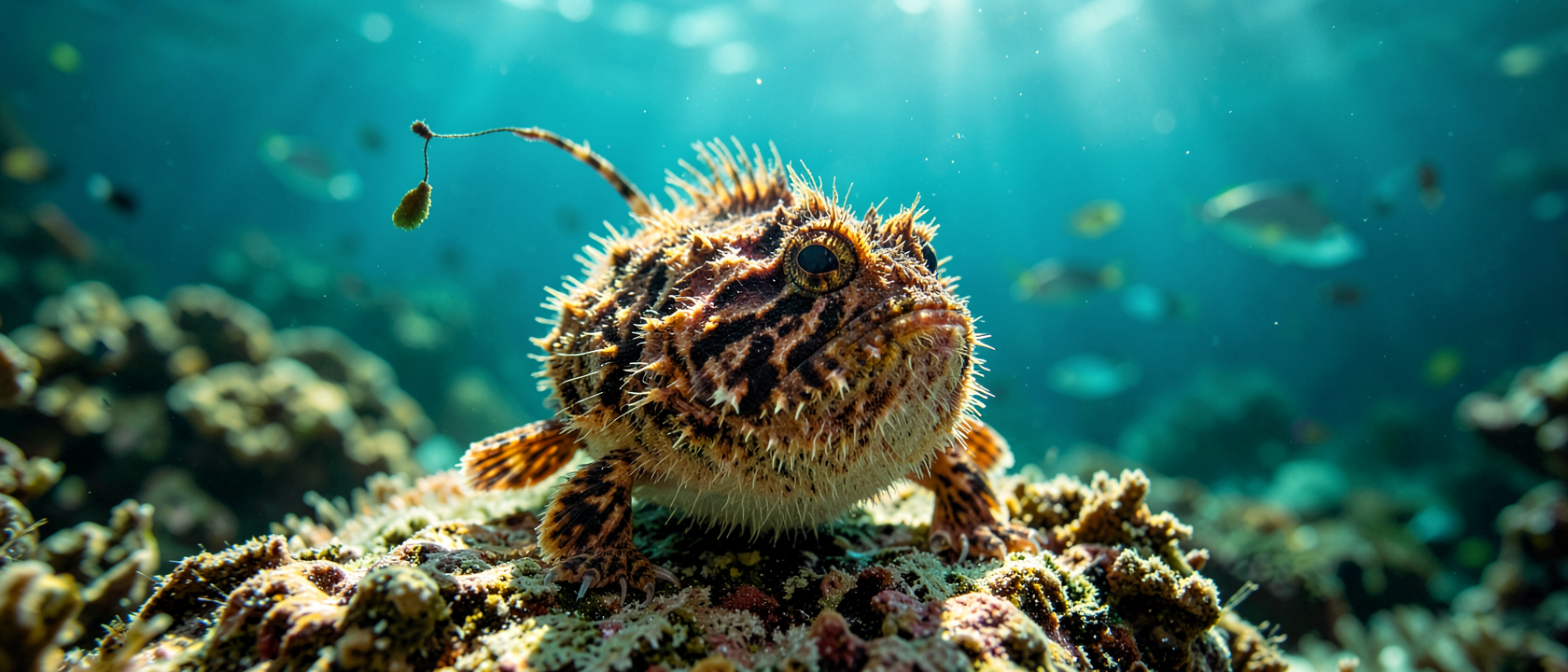
A fishing rod that rewired the spinal cord
Vertebrate motor neurons are predictable residents. In every species studied so far, the neurons commanding each fin or limb occupy a characteristic zone in the ventral horn (the anterior region of the spinal cord’s gray matter that governs movement). Dorsal-fin neurons cluster at the ventrolateral zone, the lower-outer margin of that structure. Every dorsal fin in every fish follows that rule.
Except one.
In the striated frogfish (Antennarius striatus), the motor neurons controlling the frontmost dorsal fin do not sit at the ventrolateral zone. A team led by Professor Naoyuki Yamamoto at Nagoya University’s Graduate School of Bioagricultural Sciences injected neural tracers into the muscles of that fin and followed the signals back to their source. The signals terminated at the dorsolateral zone, the upper-outer margin of the ventral horn: a location where no dorsal-fin motor neurons have ever been found in any vertebrate. Hagio, Nishino, Miyake, Sato, Sawada, Nakayama, and Yamamoto published the results in the Journal of Comparative Neurology (vol. 532, issue 10, 2024) and named the misaddressed cells “fishing motor neurons.”
The striated frogfish’s frontmost dorsal fin is the illicium: a slender, flexible rod tipped with a fleshy, worm-like lure called the esca. The fish waves the illicium above its own mouth to coax prey into range, then engulfs them. The motor neurons running that performance sit nowhere near the neurons for any other fin in the same fish, and nowhere near the corresponding neurons in any fish whose first dorsal fin is not a lure.
How a fin becomes a fishing rod
The striated frogfish has four dorsal fins. The second, third, and fourth are unremarkable: they sit along the back, contribute to posture and slow locomotion, and their motor neurons occupy the ventrolateral zone exactly as expected. The first has been repurposed entirely into the illicium, extended forward independently of all other fins to dangle the esca in front of the fish’s own mouth.
The Yamamoto team’s tracer work showed that this functional independence has a direct anatomical correlate: the neurons commanding the illicium occupy a different address in the spinal cord from the neurons commanding everything else dorsal. The circuitry of the lure is physically separate from the circuitry of ordinary fin control.
Once prey commits, the window for error is essentially zero. The striated frogfish’s buccal cavity can expand and engulf prey in under 4 milliseconds, one of the fastest feeding speeds among animals. That speed depends on the luring phase working precisely beforehand, which depends on fine, continuously independent control of the illicium. The neurological separation is the engineering reason the whole trick holds together.
The filefish holds the ancestral address
To establish that the frogfish’s dorsolateral arrangement is genuinely anomalous, Yamamoto’s team ran the same tracer experiment on the white-spotted pygmy filefish (Rudarius ercodes). The filefish’s first dorsal fin is not a lure but an erectable spine used in threat displays.
In R. ercodes, the first-dorsal-fin motor neurons sit in the ventrolateral zone, the same ancestral position occupied by all other dorsal-fin neurons in both species. The relocation documented in the frogfish is not a feature of every modified first dorsal fin. It is specific to the frogfish’s lure-waving specialization. The filefish is the control that makes that case airtight: same fin position, different function, different spinal address.
The neurons moved because the job changed
Because the filefish’s first-dorsal-fin neurons retain the ancestral ventrolateral position, the Yamamoto team interprets the frogfish’s dorsolateral cluster as the physical record of an evolutionary migration inside the central nervous system. As the fin’s function shifted from standard locomotion to precision lure-waving, the motor neuron population relocated to a zone that, as far as the published record goes, had never held a dorsal-fin neuron before.
The spatial organization of motor neuron columns in the vertebrate spinal cord has been treated as a deeply conserved ground plan: forelimb neurons here, hindlimb neurons there, pectoral-fin neurons at one characteristic address. The frogfish data put pressure on that assumption. If a single functional shift in one fin can relocate its commanding neurons to a previously unoccupied zone, the address book is more of a working draft than a finished document. Professor Yamamoto’s team draws an explicit parallel to limb motor neuron organization across vertebrates, including in humans, noting that the rules appear broadly conserved, except, demonstrably, when a fin becomes a fishing rod.
Two myths worth naming
The professional myth that dies here is the assumption that motor neuron column organization is stable enough across vertebrate lineages to be reliably predictive. The frogfish’s fishing motor neurons sitting at a dorsolateral address that nothing else dorsal occupies contradicts it at the mechanistic level.
The popular myth shapes how most readers picture this animal before they read a word about the study. The striated frogfish is an anglerfish, family Antennariidae, order Lophiiformes, and the image that word calls up is usually a deep-sea creature with a glowing lure dangling from its forehead. Frogfishes are not that animal. Antennarius striatus lives in shallow, reef-associated waters from about 33 feet (10 m) to approximately 719 feet (219 m) depth, mostly shallower than 131 feet (40 m), across tropical and subtropical Indo-Pacific and Atlantic reefs. Its esca is fleshy and non-luminous; it attracts prey through shape and motion, not light. The bioluminescent lure biology, powered by symbiotic bacteria, belongs to deep-sea ceratioid anglerfish, a different suborder entirely. The two animals are related the way a bat and a hummingbird are related: same broad group, very different machinery.
The machinery that moved the neurons is entirely unknown
Hagio, Nishino, Miyake, Sato, Sawada, Nakayama, and Yamamoto are explicit about where the 2024 paper ends. The developmental and genetic mechanisms that caused the fishing motor neurons to occupy the dorsolateral zone remain completely uncharacterized. Whether changes in axon guidance cues, transcription factor expression, or some other process drove the migration has not been established. The afferent pathways delivering sensory input to those neurons, and the descending motor commands that coordinate illicium movement with the rest of the fish’s behavior, have not been mapped. Whether other lophiiform teleosts with a functional illicium share the same dorsolateral arrangement, or whether this is specific to Antennarius striatus, is also unresolved and flagged by the team as the next comparison to run.